Cauda Equina Syndrome Mri
Cauda equina syndrome mri. The clinical and radiological findings in a patient with only a 3-year history in whom the onset of joint and neurological symptoms was apparently simultaneous are presented. The timing of surgery in cauda equina syndrome due to prolapsed intervertebral disc remains controversial. It is a time-sensitive syndrome requiring rapid and effective assessment diagnosis and treatment if.
Cauda equina syndrome incomplete CESI or CES with retention is diagnosed if the patient has clinical features of CES and radiological evidence of compression of the cauda equina on MRI. In this study we assess the ability of neurosurgical residents to predict on clinical grounds in which patients with cauda equina syndrome CES this was due to prolapsed intervertebral disc thereby justifying a request for urgent MR imaging. This syndrome has a complex spectrum of signs and symptoms.
Cauda Equina Syndrome is the signs and symptoms when the nerves of the Cauda Equina are compressed. An MRI scan must be carried out on an emergency basis because cauda equina syndrome. The cauda equina syndrome is a rare but well-recognised complication of longstanding ankylosing spondylitis usually presenting when the joint disease is quiescent.
If your doctor has suspicions of CES then in some causes it can be confirmed with a MRI scan. These symptoms should prompt medical practitioners to suspect cauda equina syndrome. CES is a neurosurgical emergency and patients undergo surgical decompression.
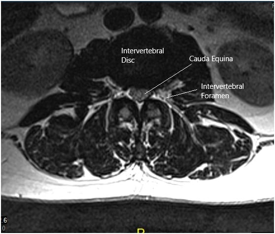
Cauda equina syndrome CES is a devastating condition for quality of life which may lead to permanent loss of bladder bowel and sexual function with associated neurological pain affecting the patient their family and relationships. Click here for Patient Education. The cauda equina is formed by the nerve roots caudal to the level of the conus medullaris.
A patient should therefore be sent for an emergency MRI scan meaning within around four hours of presenting to hospital. CT percentage thecal sac effacement of 50 predicts significant spinal stenosis on MR imaging in patients with clinically suspected cauda equina syndrome. The best way to assess the cauda equina is with MRI of the lumbar spine with and without contrast medium.
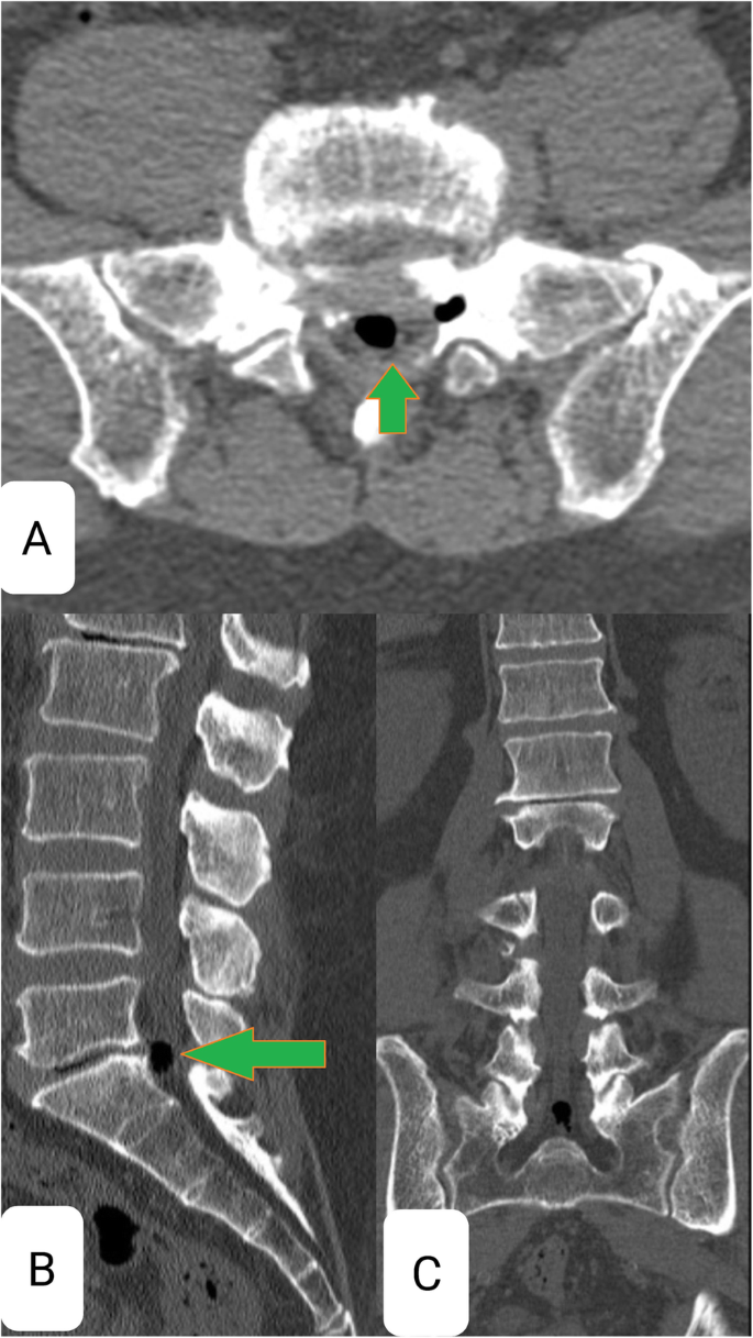
If cauda equina syndrome is possible immediately do MRI or if it is not available do CT myelography. Body of S1 vertebra.
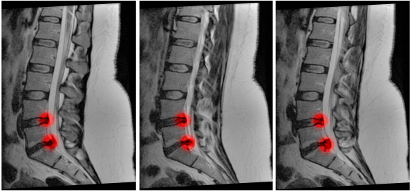
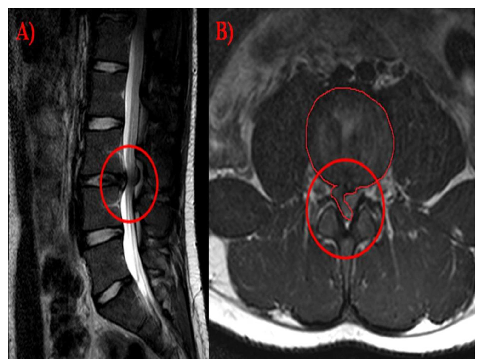
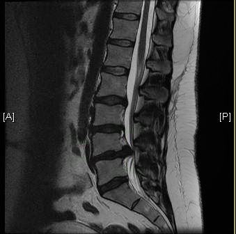
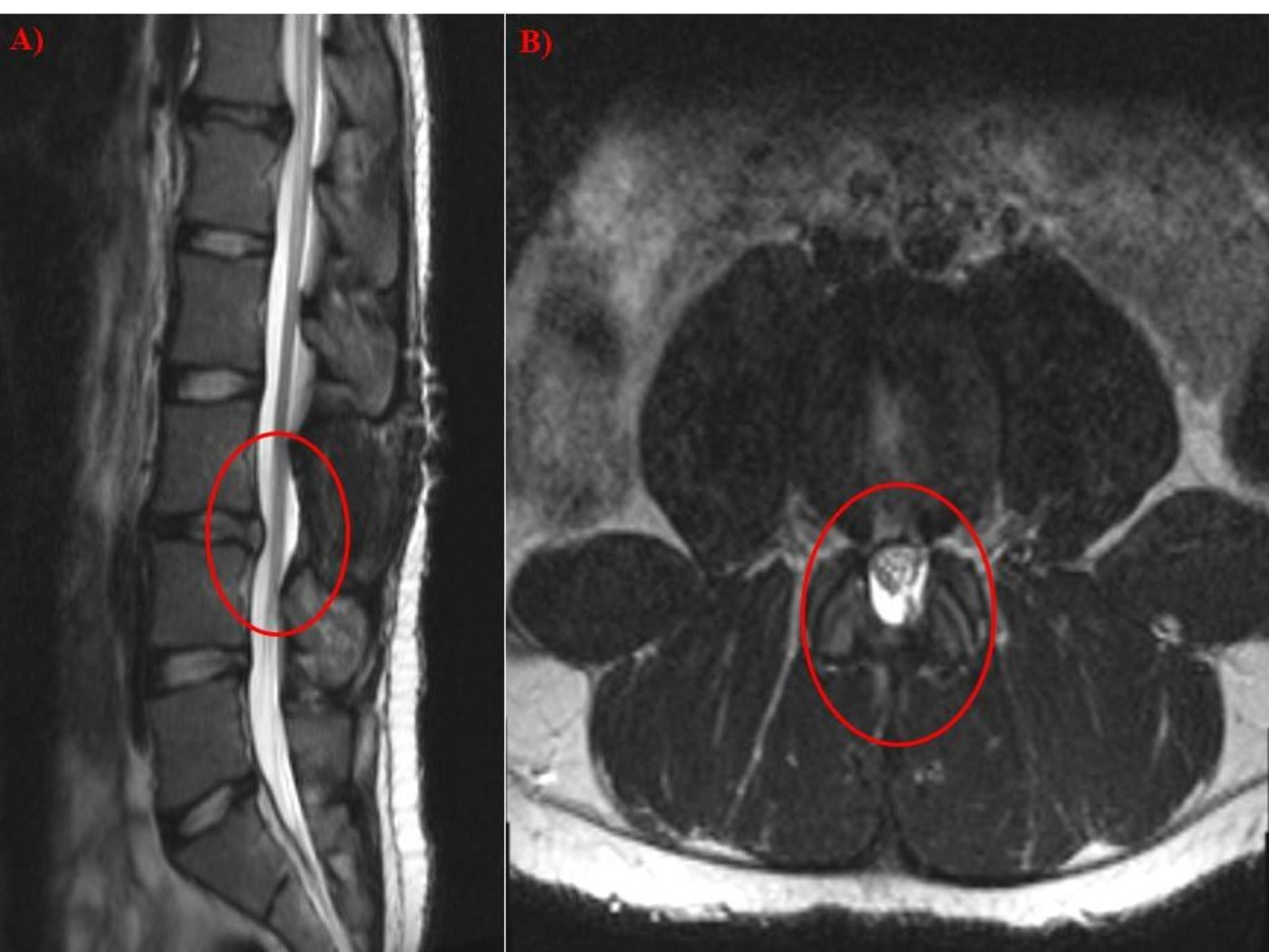
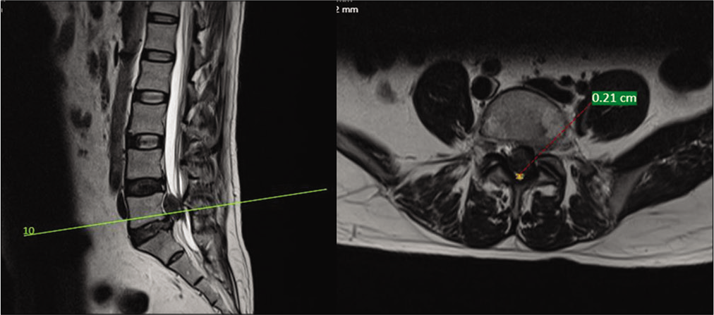
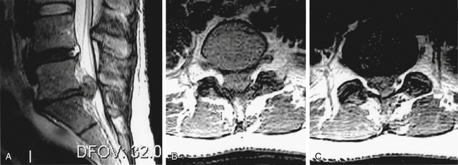
MRI scan for cauda equina syndrome.
The cauda equina syndrome is a rare but well-recognised complication of longstanding ankylosing spondylitis usually presenting when the joint disease is quiescent. Cauda Equina Syndrome is the signs and symptoms when the nerves of the Cauda Equina are compressed. MRI scan for cauda equina syndrome. Body of S1 vertebra. Of a patient with cauda equina syndrome showing a large irregular disc herniation arrow occupying most of the vertebral canal. CT percentage thecal sac effacement of. The cauda equina syndrome can result from any lesion that compresses the cauda equina and causes a dysfunction of multiple lumbar and sacral nerve roots. CES can lead to incontinence and even permanent paralysis. Whilst the Cauda Equina are protected by the vertebrae the back bone.
A patient should therefore be sent for an emergency MRI scan meaning within around four hours of presenting to hospital. Cauda equina syndrome CES is a devastating condition for quality of life which may lead to permanent loss of bladder bowel and sexual function with associated neurological pain affecting the patient their family and relationships. This is the Professional Version. Of a patient with cauda equina syndrome showing a large irregular disc herniation arrow occupying most of the vertebral canal. Cauda Equina Syndrome is the signs and symptoms when the nerves of the Cauda Equina are compressed. By contrast a cross sectional MRI view at L5S1 in a patient without cauda equina syndrome showing an unobstructed vertebral canal arrows from top down. Cauda equina syndrome CES occurs when the nerve roots of the cauda equina are compressed and disrupt motor and sensory function to the lower extremities and bladder.














Post a Comment for "Cauda Equina Syndrome Mri"